What is Glaucoma?
Glaucoma is a condition when the optic nerve suffers progressive damage with loss of nerve fibres resulting in progressive loss of vision. The condition is commonly due to raised eye pressures over a long period of time.
The eye produces fluid constantly, which is drained away through a sieve like structure called the Trabecular Meshwork at the inner corner between the cornea and the iris. When the resistance within the trabecular meshwork builds up, this will cause build up of pressure inside the eyes. The pressure will cause damage to the optic nerve head (optic disc) and result in the loss of vision.
How do you know that you have glaucoma?
Most patients suffer from glaucoma do not have any symptoms. Usually the opticians detect possible glaucoma changes with raised eye pressure, suspicious optic nerve head (optic disc) or visual field loss during routine eye tests. Patients may notice reduction in vision when visual field loss becoming more advanced in both eyes.
Some patients who suffers from acute angle closure glaucoma will get symptoms with eye pain, blurred vision, headache or brow-ache and seeing 'halos' around lights at night.
Who are at risk of glaucoma?
Around 60 million people worldwide suffer from glaucoma. In UK, it has been estimated that 50% of people with glaucoma are not diagnosed.
The following groups of people have increased risks of glaucoma:
- Older age (>50 yr old)
- Family history of glaucoma (especially in first degree relatives)
- African Caribbean ethnicity
- Extreme short and long sighted people
- Diabetic patients with poor sugar control
How is glaucoma monitored?
Glaucoma is mainly monitored by the following tests:
Eye pressure measurement
Normal eye pressure is between 8 to 21 mmHg. If the eye pressure is persistently raised above 21 mmHg, there is an increase risk of glaucoma. The higher the pressure, the higher the risk of glaucoma. In some patients, however, even the eye pressure are within normal range, they can still develop glaucoma because their optic nerves are more vulnerable and require a much lower eye pressure to reduce the risk of further glaucoma damage.
The eye pressure is commonly measured by an applanation method. After instilling anaesthetic eye drops, a small Goldmann-type applanation tonometer is commonly used with the slit-lamp to measure the eye pressure. A newer rebound tonometer (iCare) can be used when patients cannot tolerate the applanation method e.g. children.
Eye pressure day-time phasing is performed when the glaucoma specialist suspects that the patients eye pressures are fluctuating widely throughout the day, which may be causing optic nerve damage without demonstrating raised eye pressure during serial measurements in the clinic. The patient will require to attend the out-patient department for whole day eye pressure testing every 2 hours between 9:00 am to 5:00 pm.
Visual field test
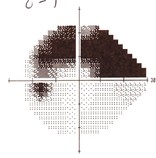
Visual field test is a test of sensitivity of the central and periperal vision. The test involves sitting in front of a dome-shaped machine. Light spots of different brightness are shined on the inner surface of the dome. You are instructed to look straight ahead at a target and press a button when you can see the light spots against a bright background.
The picture is showing a typical visual field defect (black area) due to glaucoma damage. At the early stage in glaucoma, patients may not notice the visual field defects because the other eye tends to compensate for the loss vision in some area. However, when the disease progresses, the visual fields loss in both eyes may fall on the same area and patients may start to notice that they are not seeing well in some area or objects or cars appear unexpectedly into the remaining centre of vision.
Driving can become difficult when there is significant visual field loss in both eyes. Patients must inform DVLA when they are diagnosed of suffering from glaucoma. Patients will need to complete a V1 form and DVLA will arrange a visual field test for you to be done with both eyes open. The DVLA will make decision on whether you meet the visual standard for driving base on the field test results.
Optic nerve head (Optic disc)
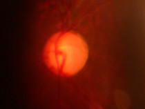
Optic disc is the optic nerve head that we can see inside the eye with a special focusing lens. In normal people, there is a small depression on the optic disc called the optic disc cup. This is normally occupying 30% of the optuc disc area and the doctor normally measure the vertical cup-to-disc ratio during routine examination. Normal cup-disc ratio is about 0.3 to 0.5 depending on the size of the optic discs. When the cup-disc ratio is more than 0.6, this is generally considered to be abnormal and require further investigation. When this is coupled with raised eye pressure, then this is considered to be glaucoma changes on the optic discs that needs treatment to lower the eye pressure. Without treatment, the optic nerve damage will continue and the cup-disc ratio will gradually enlarged over time with result of visual loss. Some patients can develop glaucome-type optic disc changes without raised eye pressure, this is consider to be Normal Pressure Glaucoma meaning that the optic nerve damage happens even when the eye pressures are within normal limits, therefore, treatment or surgery may be require to achieve lower eye pressure to protect the optic nerve.
Optic disc laser imaging
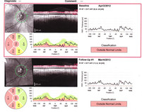
Optic disc imaging is a laser scan that can measure the thickness of nerve fibre layer surrounding the optic discs. The latest Spectral Domain Optical Coherence Tomography (SD-OCT) can accurately measure the thickness of the nerve fibre layer in micrometers. The latest eye tracking technologies allow accurate scanning of the same locations as in the previous scans, which allows accurate comparison of nerve fibre layer thickness for up to 5% of changes can be detected accurately.
The picture is showing an OCT scan of an optic nerve about 1 year apart demonstrating minimal changes of the nerve fibre layer thickness.


